Research Use Only. The information presented here is for scientific and educational purposes. These compounds are not intended for human consumption, self-administration, or therapeutic use.
Introduction
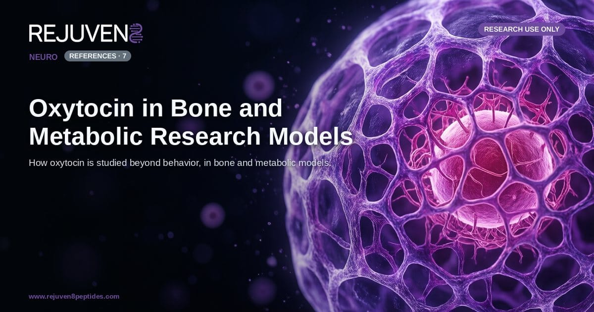
Oxytocin is most commonly described as a nonapeptide signaling molecule tied to central and social behavior, yet a substantial preclinical literature examines it in an entirely different context: the skeleton and whole-body energy balance. Research groups have reported that the oxytocin receptor (OXTR), a class A G-protein-coupled receptor, is expressed not only in the brain but also on bone cells and adipocytes. That peripheral distribution is the starting point for a body of work that studies oxytocin as a candidate regulator of osteoblast activity and metabolic phenotype in cells and animals.
This article surveys that specific literature. The focus is deliberately narrow: osteoblast biology, skeletal phenotyping in genetic models, and energy-balance readouts in rodents. This peripheral, tissue-level story is distinct from the receptor-and-behavior mechanism covered elsewhere. Every observation summarized here is drawn from in vitro systems and animal models and is presented for research use only, not for human consumption. No claim of human dosing, efficacy, or benefit is made or implied.
Oxytocin as a Nonapeptide: Molecular Identity
Before reviewing the model systems, it helps to fix the identity of the molecule being studied. Oxytocin is a small, cyclic peptide. Its compact structure and defined disulfide topology make it a well-characterized reference compound in laboratory work, and the same molecule appears across the bone and metabolic studies discussed below. Investigators sourcing a research-grade Oxytocin reference typically confirm identity and purity against a certificate of analysis before use.
The values below reflect commonly reported laboratory attributes and are provided for comparison only.
| Attribute | Commonly Reported Value |
|---|---|
| Class | Cyclic neurohypophysial nonapeptide |
| Amino acid sequence | Cys-Tyr-Ile-Gln-Asn-Cys-Pro-Leu-Gly-NH2 |
| Sequence length | 9 residues (nonapeptide) |
| Molecular formula | C43H66N12O12S2 |
| Molecular weight | approximately 1007 g/mol |
| Structural feature | Intramolecular disulfide bond (Cys1 to Cys6) |
| Receptor target | Oxytocin receptor (OXTR), a class A GPCR |
Oxytocin differs from the related peptide vasopressin by only two amino acid residues, which is one reason receptor selectivity and possible cross-reactivity are recurring themes in the primary literature. The disulfide-bridged ring is central to how the peptide engages OXTR, and it is the reason handling protocols emphasize careful reconstitution and storage.
Oxytocin in Bone Research Models
The skeletal literature treats oxytocin as a possible local and systemic signal acting on the two principal bone cell types: bone-forming osteoblasts and bone-resorbing osteoclasts. Because both lineages have been reported to express OXTR in culture, oxytocin is studied as a candidate modulator of the balance between bone formation and resorption in preclinical systems.
Osteoblast differentiation and mineralization in vitro
A large share of the bone work is cell-based. Using primary osteoblasts and osteoblast-like cell lines, investigators examine whether exposure to oxytocin is associated with changes in differentiation markers such as RUNX2, alkaline phosphatase, osteocalcin, and collagen expression, along with the formation of mineralized nodules. These assays measure osteoblast behavior in a dish and are used to generate hypotheses about how OXTR signaling might intersect with established bone-formation pathways. They are not statements about any living organism beyond the culture system.
Receptor and ligand knockout skeletal phenotypes
A second line of evidence comes from genetic mouse models. Studies of OXTR-null and oxytocin-deficient mice have reported altered bone mass phenotypes relative to wild-type littermates, which is the basis for describing oxytocin signaling as relevant to skeletal homeostasis in that species. Phenotyping in these models typically relies on micro-computed tomography, histomorphometry, and mechanical testing of excised bones to quantify trabecular and cortical parameters.
Ovariectomized rodent models of bone loss
The ovariectomized (OVX) rodent is a standard preclinical model of estrogen-deficient bone loss. Within this framework, oxytocin has been investigated for its association with the anabolic action of estrogen on the skeleton and for its interaction with bone marrow signaling. Because estrogen status and oxytocin signaling appear linked in these animals, the OVX model is frequently used to probe that relationship under controlled conditions. All such findings are confined to the animal models in which they were observed.
Oxytocin in Metabolic Research Models
Parallel to the bone literature is a body of work on energy balance. Here oxytocin is studied primarily in rodents and in adipose cell cultures, with readouts centered on food intake, body composition, and lipid and glucose handling. As with the skeletal studies, these are model-system observations rather than human findings.
Diet-induced obesity models
The diet-induced obesity (DIO) rodent is the workhorse of this field. In DIO rats and mice, researchers examine associations between oxytocin administration and measures such as caloric intake, body weight trajectory, and adipose depot size. These experiments are designed to characterize the peptide within a defined animal paradigm and are interpreted strictly as preclinical data.
Adipocyte receptor expression and lipid handling
At the cellular level, adipocytes and preadipocyte lines are used to study whether OXTR is expressed on fat cells and how oxytocin exposure relates to markers of lipolysis, lipid uptake, and adipogenic differentiation. This in vitro work complements the whole-animal studies by isolating tissue-level responses from systemic effects.
Glucose handling and bone-energy crosstalk
Some studies connect the two themes of this article. Because osteoblasts secrete factors implicated in energy metabolism, oxytocin sits at a plausible intersection of skeletal and metabolic biology, and glucose-tolerance readouts are sometimes paired with bone endpoints in the same rodent cohort. The comparison table below summarizes how the principal model systems differ. These values reflect commonly reported laboratory attributes and are provided for comparison only.
| Model system | Primary tissue focus | Typical research readouts |
|---|---|---|
| Osteoblast cell culture | Bone formation | RUNX2, osteocalcin, mineralized nodules |
| OXTR-null / OT-null mice | Skeletal phenotype | Micro-CT, histomorphometry, bone strength |
| Ovariectomized rodent | Estrogen-deficient bone loss | Trabecular bone volume, marrow signaling |
| Diet-induced obesity rodent | Whole-body metabolism | Food intake, body weight, adiposity |
| Adipocyte cell culture | Lipid metabolism | OXTR expression, lipolysis, adipogenesis |
Interpreting the Bone and Metabolic Literature
Several caveats accompany this research area. Findings are species-specific and route-specific, and results in cultured cells do not necessarily translate to intact animals, let alone across species. The close structural relationship between oxytocin and vasopressin means receptor cross-reactivity must be controlled for when interpreting signaling data. Dose, formulation, and peptide integrity all influence outcomes, so reproducibility depends heavily on well-characterized reference material.
For that reason, laboratories working in this space place weight on documentation and sourcing. Reviewing a certificate of analysis to confirm identity and purity, applying a structured approach to supplier vetting, and cross-checking published third-party certificates are standard steps before a compound enters a study. Investigators comparing peptides across projects often start from a broad research peptide catalog and then narrow to a single well-documented lot. None of these practices change the fundamental status of the material: it is intended for research use only, not for human consumption.
Frequently Asked Questions
What is oxytocin studied for in bone research?
In preclinical bone research, oxytocin is investigated as a signaling peptide that may act on bone-forming osteoblasts and bone-resorbing osteoclasts, both of which have been reported to express the oxytocin receptor (OXTR). Cell and animal studies examine its association with osteoblast differentiation and mineralization. These are laboratory findings only and do not describe any human effect.
Do osteoblasts express oxytocin receptors?
Multiple in vitro studies report OXTR expression on cultured osteoblasts and osteoclast precursors, which is the rationale for studying oxytocin as a local regulator of bone cell activity in research models. This describes receptor biology observed in cells rather than any clinical outcome.
How is oxytocin investigated in metabolic research models?
Metabolic studies typically use rodent models such as diet-induced obesity, examining associations between oxytocin administration and readouts like food intake, body weight, adiposity, and glucose handling in the animals. Adipocyte cell cultures are also used to study receptor expression and lipid metabolism. All of this work is confined to model systems.
Is the oxytocin in these studies the same molecule as the pituitary hormone?
Yes, the compound studied is the same cyclic nonapeptide, oxytocin, with the sequence Cys-Tyr-Ile-Gln-Asn-Cys-Pro-Leu-Gly-NH2. Bone and metabolic research examines peripheral actions of this peptide that are distinct from its classical central and behavioral roles.
What is the molecular weight of oxytocin?
Oxytocin has a molecular weight of approximately 1007 g/mol and a molecular formula of C43H66N12O12S2. It is a nine-amino-acid peptide featuring an intramolecular disulfide bond between two cysteine residues.
Can oxytocin be used outside the laboratory?
No. All materials referenced here are for research use only, not for human consumption. The literature described involves in vitro systems and animal models, and none of it constitutes evidence of human safety, dosing, or benefit.
References and Further Reading
- Tamma R, Colaianni G, and colleagues, work describing oxytocin as an anabolic signal in bone (Proceedings of the National Academy of Sciences, 2009). PubMed: oxytocin anabolic bone
- Colaianni G and colleagues, studies on bone marrow oxytocin and the action of estrogen on the skeleton. PubMed: oxytocin bone marrow estrogen
- Elabd C and colleagues, work on oxytocin and the osteoblast to adipocyte balance in aging models. PubMed: oxytocin osteoblast adipocyte
- Ovariectomized rodent studies of estrogen-deficient bone loss and oxytocin signaling. PubMed: oxytocin ovariectomized bone
- Deblon N and colleagues, anti-obesity effects of oxytocin in diet-induced obese rodents (2011). PubMed: oxytocin diet-induced obesity
- Blevins JE and colleagues, reviews on oxytocin and energy balance in animal models. PubMed: oxytocin energy balance
- Reports on oxytocin receptor (OXTR) expression in skeletal and adipose tissue. PubMed: oxytocin receptor bone metabolism