Research Use Only. The information presented here is for scientific and educational purposes. These compounds are not intended for human consumption, self-administration, or therapeutic use.
Introduction
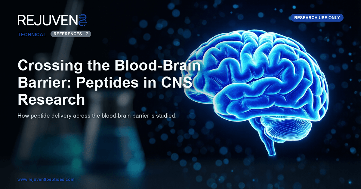
The blood-brain barrier (BBB) is one of the most selective interfaces in mammalian physiology and a central obstacle in preclinical central nervous system (CNS) research. Formed by specialized brain microvascular endothelial cells joined by continuous tight junctions, it restricts the passage of most large or polar molecules from blood into brain tissue. Peptides, which are typically hydrophilic and enzymatically labile, are among the classes most affected, so how blood-brain barrier peptides cross this endothelium is a recurring question in neuropharmacology and molecular delivery.
This article surveys how peptide penetration and transport across the blood-brain barrier are investigated in preclinical and in-vitro settings. It covers the biological routes that have been characterized, including receptor-mediated transcytosis and adsorptive mechanisms, the shuttle-peptide design strategies used to exploit them, and the models used to quantify permeability. All material is provided for research use only, not for human consumption, and no clinical, dosing, or therapeutic conclusions are drawn.
Blood-Brain Barrier Peptides: What Limits Their Entry
The neurovascular unit
The barrier is best understood as part of a neurovascular unit, a functional assembly of endothelial cells, pericytes, astrocyte end-feet, and the surrounding basement membrane. Endothelial cells here differ markedly from peripheral capillaries: they lack fenestrations, show minimal pinocytotic activity, and are sealed by tight junction complexes built from claudins, occludin, and zonula occludens proteins. This architecture forces molecules through the cells (the transcellular route) rather than between them, effectively closing the paracellular pathway to circulating peptides.
Barriers to peptide entry
Several properties keep most peptides out of the brain parenchyma. Molecular size and high polar surface area limit passive diffusion, because hydrophilic peptides partition poorly into the lipid membrane. Enzymatic degradation by peptidases in plasma and at the endothelial surface shortens circulating half-life. Efflux transporters such as P-glycoprotein (P-gp) and other ABC family members actively return many substrates to the blood. Together these factors keep the fraction reaching the CNS very low in animal models, which is why targeted transport strategies are studied intensively.
Routes of Transport Studied Across the Barrier
Researchers studying blood-brain barrier peptides distinguish several transport routes, each characterized by different cargo, kinetics, and specificity.
Passive and carrier-mediated routes
A few lipophilic molecules cross by passive transcellular diffusion, but this route is largely closed to typical peptides. Carrier-mediated transport (CMT) uses solute carriers, for example those for glucose or specific amino acids, and generally accommodates only small, structurally defined substrates. Some short peptides and peptidomimetics have been engineered to resemble these carrier substrates.
Receptor-mediated transcytosis
Receptor-mediated transcytosis (RMT) is the most studied route for larger cargo. Endogenous ligands bind receptors on the luminal endothelial surface, are internalized in vesicles, and are released on the brain side. Frequently studied systems include the transferrin receptor (TfR), the insulin receptor, and low-density lipoprotein receptor-related protein 1 (LRP1). Ligands, engineered peptides, or antibodies to these receptors act as targeting moieties.
Adsorptive-mediated transcytosis
Adsorptive-mediated transcytosis (AMT) is triggered by electrostatic interaction between cationic molecules and the negatively charged endothelial glycocalyx. Because it requires no specific receptor, AMT is the route most associated with cationic cell-penetrating peptides. It is generally less specific than RMT, a trade-off researchers weigh when designing delivery vectors.
The following comparison reflects commonly reported laboratory attributes and is provided for comparison only.
| Transport route | Physical basis | Typical cargo studied | Representative target or example |
|---|---|---|---|
| Passive diffusion | Lipid solubility | Small lipophilic molecules | Not favorable for most peptides |
| Carrier-mediated transport | Solute carrier proteins | Small substrates, peptidomimetics | Amino acid and glucose carriers |
| Receptor-mediated transcytosis | Ligand-receptor vesicular transport | Larger peptides, proteins, conjugates | Transferrin receptor, insulin receptor, LRP1 |
| Adsorptive-mediated transcytosis | Electrostatic (cationic) binding | Cationic peptides and conjugates | Cell-penetrating peptides (TAT, penetratin) |
Shuttle Peptides and Molecular Design Strategies
Once a transport route is identified, the question becomes how to build a peptide that engages it. Two broad approaches dominate the literature: dedicated shuttle sequences and chemical modification of the backbone.
BBB shuttle peptides
A BBB shuttle peptide is a sequence designed to ferry an attached cargo across the endothelium, usually by engaging an RMT receptor. Angiopep-2, a 19-residue peptide derived from a Kunitz domain and targeting LRP1, is among the most cited examples in preclinical delivery research. Other shuttles come from phage-display screening or from natural venom and protein sequences. These constructs are studied as vectors: the shuttle is not the payload but the transport component.
Cell-penetrating peptides
Cell-penetrating peptides (CPPs) such as the HIV-derived TAT peptide and penetratin are short, often arginine-rich sequences that promote cellular uptake, partly through adsorptive-mediated transcytosis. In BBB research they are evaluated both alone and fused to cargo, though their relatively low tissue specificity is a recognized limitation.
Chemical modification strategies
Independent of the transport route, medicinal chemists modify peptide backbones to improve stability and permeability. Approaches studied in the literature include cyclization, incorporation of D-amino acids to resist peptidase cleavage, N-methylation, glycosylation, lipidation, and PEGylation. Small nootropic research peptides such as Semax and Selank, and the sleep-associated peptide DSIP, are frequently referenced in discussions of central activity because of their compact size and reported effects in animal models.
The values below reflect commonly reported laboratory attributes and are provided for comparison only.
| Peptide | Sequence length | Approx. molecular weight | Class or studied context |
|---|---|---|---|
| Semax | 7 residues | ~813 Da | ACTH(4-10) analog, nootropic research |
| Selank | 7 residues | ~751 Da | Tuftsin analog, anxiolytic research |
| DSIP | 9 residues | ~849 Da | Delta sleep-inducing peptide |
| Angiopep-2 | 19 residues | ~2400 Da | LRP1-targeting BBB shuttle |
| TAT (47-57) | 11 residues | ~1560 Da | Cationic cell-penetrating peptide |
In-Vitro and In-Vivo Models Used to Study BBB Penetration
No single model captures the whole barrier, so the penetration of blood-brain barrier peptides is assessed with several methods.
In-vitro barrier models
The workhorse in-vitro system is a monolayer of brain endothelial cells grown on a porous Transwell insert, which separates an apical (blood-side) compartment from a basolateral (brain-side) compartment. Barrier integrity is monitored by transendothelial electrical resistance (TEER), and permeability is expressed as an apparent permeability coefficient (Papp). Co-culture with pericytes or astrocytes, and newer microfluidic BBB-on-chip platforms, reproduce more of the neurovascular unit and its shear-flow environment. Results depend heavily on cell source and documentation, as covered in this certificate of analysis guide.
In-situ and in-vivo methods
In-situ brain perfusion measures uptake directly from a controlled vascular input in rodents. Microdialysis samples free compound in the brain interstitial fluid over time, while cerebrospinal fluid sampling gives an indirect readout of central exposure. Radiolabeling and imaging methods such as positron emission tomography (PET) track distribution quantitatively. Because models differ in what they capture, results are interpreted across several methods, not a single assay. Compounds used in this work are drawn from a research peptide catalog and handled strictly as laboratory reagents.
Frequently Asked Questions
Can peptides cross the blood-brain barrier?
Most peptides cross the intact blood-brain barrier only in small amounts, because their size, polarity, and susceptibility to enzymatic degradation limit passive entry. Preclinical research therefore focuses on transport routes such as receptor-mediated transcytosis and on engineered shuttle peptides that can carry cargo across brain endothelial cells in laboratory models.
What is receptor-mediated transcytosis?
Receptor-mediated transcytosis (RMT) is a vesicular transport process in which a molecule binds a receptor on the blood-facing surface of brain endothelial cells, is internalized in vesicles, and is released on the brain side. Transferrin, insulin, and LRP1 receptors are among the most commonly studied RMT targets in delivery research.
What are BBB shuttle peptides?
BBB shuttle peptides are short sequences designed to transport an attached cargo across the barrier, usually by engaging a transcytosis receptor. Angiopep-2, which targets LRP1, is a widely cited example in preclinical delivery studies, where the shuttle acts as a transport vector rather than as the active payload.
How is blood-brain barrier penetration measured in vitro?
A common in-vitro approach grows brain endothelial cells as a monolayer on a porous Transwell insert. Barrier tightness is monitored with transendothelial electrical resistance (TEER), and the movement of a test compound is quantified as an apparent permeability coefficient (Papp). Co-cultures and microfluidic chip models add further physiological detail.
Why is the blood-brain barrier so difficult for peptides to cross?
The barrier combines tight junctions that block the paracellular route, very low vesicular activity, peptidases at the endothelial surface and in plasma, and active efflux transporters such as P-glycoprotein. These features together keep the brain uptake of most circulating peptides very low in animal studies.
Do cell-penetrating peptides cross the blood-brain barrier?
Cationic cell-penetrating peptides such as TAT and penetratin can promote uptake across cell membranes, partly through adsorptive-mediated transcytosis. In blood-brain barrier research they are studied both alone and fused to cargo, although their limited tissue specificity is a recognized constraint.
References and Further Reading
- Banks, W. A., and coworkers. Body of work on peptide and protein transport across the blood-brain barrier. PubMed: peptide transport across the blood-brain barrier
- Pardridge, W. M. Reviews on receptor-mediated transcytosis and molecular Trojan horse strategies for CNS delivery. PubMed: receptor-mediated transcytosis
- Teixido, M., and Giralt, E. Research on the design of blood-brain barrier shuttle peptides. PubMed: blood-brain barrier shuttle peptides
- Preclinical studies of Angiopep-2 and LRP1-targeted transport vectors. PubMed: Angiopep-2 transport
- Literature on cell-penetrating peptides in brain delivery research. PubMed: cell-penetrating peptides brain delivery
- Methodological literature on in-vitro blood-brain barrier models, TEER, and permeability assays. PubMed: in vitro blood-brain barrier models
- Reviews of microfluidic blood-brain barrier organ-on-chip platforms. PubMed: blood-brain barrier on-chip models