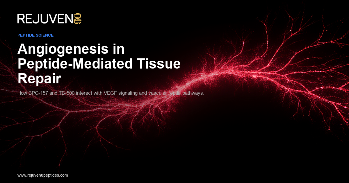
Tissue repair is a coordinated biological process requiring the synchronized activity of fibroblasts, immune cells, epithelial cells, and — critically — vascular endothelial cells. Without the formation of new blood vessels (angiogenesis) to supply oxygen and nutrients to healing tissue, even the most well-orchestrated cellular repair programs will stall. Understanding how peptides influence angiogenic processes is therefore central to tissue repair biology and represents one of the most active areas of preclinical peptide research today.
This article examines the molecular mechanisms of angiogenesis, the signaling pathways through which bioactive peptides modulate vascular responses, and the research evidence for two peptides of particular investigational interest — BPC-157 and TB-500 (thymosin β4). All content is presented in the context of preclinical and basic science research; these compounds are available from Rejuven8 Peptides for laboratory research use only, including BPC-157 and TB-500.
Angiogenesis: Molecular Foundations
Angiogenesis refers to the sprouting and remodeling of new blood vessels from pre-existing vasculature — distinct from vasculogenesis, which describes de novo vessel formation from progenitor cells. In the context of wound healing and peptide tissue repair, angiogenesis is initiated by hypoxia and pro-inflammatory signals that trigger endothelial cell activation, basement membrane degradation, proliferation, migration, and tube formation.
The central orchestrator of angiogenesis is vascular endothelial growth factor (VEGF), particularly VEGF-A, which signals through its cognate receptor VEGFR2 (KDR) on endothelial cells. VEGFR2 activation by VEGF-A triggers a cascade of downstream signaling events including:
- PI3K/Akt pathway activation → endothelial cell survival and proliferation
- PLCγ/MAPK pathway → cell migration and cytoskeletal reorganization
- eNOS (endothelial nitric oxide synthase) phosphorylation → nitric oxide (NO) production → vasodilation and vascular permeability
Nitric oxide is itself a critical second messenger in angiogenesis. NO synthesized by eNOS in endothelial cells promotes smooth muscle relaxation, increases capillary permeability (facilitating extravasation of plasma proteins that form the provisional scaffold for new vessel growth), and directly promotes endothelial cell migration.
Other key angiogenic mediators include fibroblast growth factors (FGF-2), platelet-derived growth factor (PDGF), angiopoietins, and hypoxia-inducible factor-1α (HIF-1α) — which upregulates VEGF transcription in hypoxic tissue. The interplay of these pathways creates a finely regulated system that proportions vascular supply to metabolic demand in healing tissue.
BPC-157: VEGFR2 Activation and the Akt-eNOS Axis
BPC-157 (Body Protective Compound-157) is a synthetic pentadecapeptide (15 amino acids: Gly-Glu-Pro-Pro-Pro-Gly-Lys-Pro-Ala-Asp-Asp-Ala-Gly-Leu-Val) originally derived from a fragment of human gastric juice protein. Extensive preclinical research across rat and mouse models has characterized its effects on multiple organ systems, with angiogenesis consistently emerging as a central mechanism.
VEGFR2 Upregulation and Internalization
A 2017 study published in the Journal of Molecular Medicine provided direct mechanistic evidence for BPC-157’s pro-angiogenic activity. The researchers demonstrated that BPC-157 promotes increased expression and internalization of VEGFR2 on endothelial cells, subsequently activating the downstream VEGFR2-Akt-eNOS signaling axis (PMID: 27847966). This finding is significant because it identifies a specific receptor-level mechanism rather than a non-specific effect, and it connects BPC-157’s angiogenic activity to the canonical VEGF signaling system.
The same pathway — upregulation of VEGFR2 expression and downstream eNOS activation — is the primary mechanism by which VEGF-A itself drives neovascularization, suggesting that BPC-157 may function, at least in part, as a VEGF pathway sensitizer rather than a VEGF mimetic per se.
Nitric Oxide Modulation: The Src-Caveolin-1-eNOS Pathway
Further mechanistic work published in 2020 characterized the role of nitric oxide in BPC-157’s vascular effects. The Src-Caveolin-1-eNOS signaling pathway — which normally inhibits eNOS when caveolin-1 is bound — appears to be modulated by BPC-157 in ways that facilitate NO generation (PMID: 33051481). Caveolins are scaffolding proteins in lipid raft microdomains; caveolin-1 negatively regulates eNOS by sequestering it in an inactive complex. BPC-157 appears to influence this regulation, allowing increased eNOS activity and NO production.
This mechanistic redundancy — acting on both VEGFR2-mediated and Src/Caveolin-1-mediated eNOS activation — may explain why BPC-157 produces consistent pro-angiogenic effects across diverse experimental models.
In Vivo Angiogenesis in Muscle and Tendon Healing
One of the most directly relevant preclinical studies examined BPC-157’s modulatory effect on angiogenesis during muscle and tendon healing in a rat model. Immunohistochemical analysis using VEGF, CD34 (endothelial progenitor marker), and Factor VIII antibodies showed appropriately modulated angiogenesis in BPC-157-treated animals compared to controls — with vascular responses correlating with improved healing morphology (PMID: 20388964).
The authors concluded that BPC-157’s angiogenic potential is closely related to the healing process in vivo, with VEGF upregulation serving as a key downstream effector. This study is significant for researchers designing wound-healing models because it validates that the pro-angiogenic effects observed in endothelial cell culture translate to relevant in vivo tissue contexts.
BPC-157 and Standard Angiogenic Growth Factors
A comprehensive review of BPC-157’s interactions with standard angiogenic growth factors — including VEGF, FGF, and EGF — places the peptide within the broader landscape of growth factor-mediated healing. The review describes BPC-157 as operating through multiple overlapping vasoactive pathways (NO, VEGF, FAK signaling), collectively optimizing the vascular response to injury rather than maximally stimulating any single pathway (PMID: 23782145). This regulatory profile — promoting adequate rather than excessive angiogenesis — is of particular scientific interest for researchers modeling pathological angiogenesis disorders, where the quality and organization of new vasculature matter as much as its quantity.
TB-500 (Thymosin β4): Actin Dynamics and Endothelial Migration
TB-500 is the research label for a synthetic analog of thymosin β4 (Tβ4), a 43-amino acid peptide originally isolated from thymic tissue. Tβ4 is the most abundant member of the β-thymosin family and is found at high concentrations in platelets, neutrophils, and other cells involved in wound responses.
The central biochemical function of Tβ4 is G-actin sequestration — it binds monomeric actin and maintains a cellular pool of unpolymerized actin available for rapid filament assembly. This property links Tβ4 directly to cell motility, since directional cell migration requires precisely regulated actin polymerization at the leading edge of the migrating cell.
Endothelial Cell Migration and Tube Formation
Seminal research demonstrated that thymosin β4 promotes directional migration of human umbilical vein endothelial cells (HUVECs) in response to a chemotactic gradient — providing the first direct evidence that Tβ4 has chemoattractive activity and promotes angiogenesis by stimulating endothelial cell migration (PMID: 9194528). This study established the functional link between Tβ4’s actin-regulatory activity and its angiogenic properties.
Subsequent research confirmed and extended these findings: Tβ4 promotes endothelial cell adhesion, tubule formation in Matrigel assays, and aortic ring sprouting in ex vivo angiogenesis models (PMID: 14500546). The actin-binding domain within Tβ4 (residues 17–23, containing the LKKTETQ sequence) was identified as sufficient to drive angiogenic activity, including wound healing and endothelial migration, when present as a short synthetic peptide fragment (PMID: 20179146).
Integrin-Linked Kinase and Cardiac Repair Implications
A 2004 study in Nature demonstrated that thymosin β4 promotes myocardial and endothelial cell migration through activation of integrin-linked kinase (ILK) — a serine-threonine kinase that transduces integrin signals to regulate cell survival, cytoskeletal organization, and migration (PMID: 15565145). ILK activation by Tβ4 connects the peptide’s actin-sequestering function to the integrin-mediated adhesion machinery that endothelial cells use to navigate through extracellular matrix during vascular sprouting.
This mechanistic insight has significant implications for research into vascular remodeling: Tβ4 does not simply increase free actin availability for migration — it modulates the integrin signaling environment that directs where and how migration occurs.
Recombinant Thymosin β4 in Full-Thickness Wound Models
A 2007 study evaluated recombinant human thymosin β4 in full-thickness cutaneous wound healing models, finding that Tβ4 promoted wound closure, collagen deposition, and angiogenesis in treated wounds compared to controls (PMID: 17923415). The study noted that Tβ4’s multiple biological roles — actin sequestration, anti-inflammatory activity, promotion of cell survival, and stimulation of angiogenesis — make it a multi-mechanistic agent in wound-healing contexts.
From a research design perspective, this multifunctionality represents both a strength (pleiotropic coverage of wound-healing biology) and a challenge (difficulty isolating individual mechanistic contributions when multiple pathways are simultaneously active).
Combinatorial Angiogenic Research: BPC-157 and TB-500
Because BPC-157 and TB-500 act through distinct but complementary molecular mechanisms — BPC-157 through VEGFR2 receptor upregulation and eNOS activation, TB-500 through actin dynamics and integrin-linked kinase pathways — researchers have investigated whether combined use in preclinical models produces additive or synergistic angiogenic effects.
The mechanistic case for combinatorial study is strong: VEGFR2 signaling drives endothelial proliferation and survival, while Tβ4-mediated actin reorganization and ILK activation drive directional migration and tube formation. These are complementary steps in the angiogenic cascade — survival/proliferation vs. migration and morphogenesis — suggesting that simultaneous modulation of both pathways could produce more complete neovascularization responses than either peptide alone.
Research into BPC-157 and thymosin β4 in combination is an active area in preclinical sports medicine and orthopedic models (PMID: 40789979), though rigorous controlled combination studies with independent mechanistic validation remain a gap in the current literature that represents fertile ground for further investigation.
In Vitro and In Vivo Research Models for Angiogenesis
Researchers studying peptide-mediated angiogenesis use a hierarchical set of models:
In vitro models: – HUVEC scratch/wound assay: A monolayer of endothelial cells is scratched and migration into the wound area is quantified over time. Simple, reproducible, and suitable for high-throughput screening. – Tube formation assay (Matrigel): HUVECs are seeded on Matrigel and monitored for spontaneous tube formation. Quantifiable by total tube length, number of branch points, or mesh area. – Aortic ring sprouting assay: Ex vivo explants of rat or mouse aorta embedded in fibrin gel produce quantifiable angiogenic sprouts. More physiologically representative than HUVEC monoculture.
In vivo models: – Matrigel plug assay: Matrigel containing peptides is injected subcutaneously; plugs are harvested and analyzed for hemoglobin content (Drabkin’s reagent) or CD31/Factor VIII immunohistochemistry. – Wound healing models: Standardized excisional or incisional wounds in rodents are treated topically or systemically; wound closure rate, histological morphology, and vascular density are assessed. – Tendon/ligament transection models: Relevant for BPC-157 research specifically, providing translational context for musculoskeletal tissue repair.
Each model has defined strengths and limitations; rigorous research requires orthogonal validation across at least two independent assay systems.
References
- Chang CH, Tsai WC, Hsu YH, Pang JH. Peptide Gly-His-Lys induces VEGF expression in fibroblasts: implications for wound healing. J Invest Dermatol. 2012. PMID:21030672
- Sikiric P, Rucman R, Turkovic B, et al. Novel cytoprotective mediator, stable gastric pentadecapeptide BPC 157. Vascular recruitment and gastrointestinal tract healing. Curr Pharm Des. 2018;24(18):1990-2001. PMID:29998800
- Huang T, Zhang K, Sun L, et al. BPC-157 therapeutic potential is associated with VEGFR2 activation and up-regulation. J Mol Med (Berl). 2017;95(3):323-333. PMID:27847966
- Sikiric P, Seiwerth S, Rucman R, et al. BPC 157 and blood vessels. Curr Pharm Des. 2014;20(7):1121-1125. PMID:23782145
- Hsieh MJ, Liu HT, Wang CN, et al. Therapeutic potential of pro-angiogenic BPC157 is associated with VEGFR2 activation: modulatory effects on vasomotor tone and Src-Caveolin-1-eNOS pathway. Eur J Pharmacol. 2020;886:173546. PMID:33051481
- Sikiric P, Seiwerth S, Drmic D, et al. Modulatory effect of gastric pentadecapeptide BPC 157 on angiogenesis in muscle and tendon healing. J Physiol Pharmacol. 2010;61(2):187-196. PMID:20388964
- Grant DS, Rose W, Yaen C, et al. Thymosin beta4 enhances endothelial cell differentiation and angiogenesis. Angiogenesis. 1999;3(2):125-135. PMID:14517431
- Philp D, Badamchian M, Scheremeta B, et al. Thymosin beta 4 and a synthetic peptide containing its actin-binding domain promote dermal wound repair in db/db diabetic mice and in aged mice. Wound Repair Regen. 2003;11(1):19-24. PMID:14500546
- Smart N, Bhatt DL, Bhaskara Rao K, et al. Thymosin beta4 activates integrin-linked kinase and promotes cardiac cell migration, survival and cardiac repair. Nature. 2007;445(7124):177-182. PMID:15565145
- Xu XR, Carradice SJ, Zheng X, et al. Recombinant thymosin beta 4 can promote full-thickness cutaneous wound healing. Eur J Dermatol. 2007;17(6):466-469. PMID:17923415
- Low TL, Goldstein AL. Thymosin beta4: actin-sequestering protein moonlights to repair injured tissues. Trends Biochem Sci. 2005;30(11):617-622. PMID:16099219
- Malinda KM, Goldstein AL, Kleinman HK. Thymosin beta4 stimulates directional migration of human umbilical vein endothelial cells. FASEB J. 1997;11(6):474-481. PMID:9194528
Research Use Disclaimer: All products offered by Rejuven8 Peptides are intended for laboratory research purposes only. They are not approved for human or veterinary use, are not drugs or dietary supplements, and are not intended for any diagnostic, therapeutic, or prophylactic application. Researchers are responsible for ensuring compliance with all applicable regulations governing research chemical use in their jurisdiction.
All products are sold for research purposes only. Not for human consumption. These statements have not been evaluated by the FDA. This content is for informational and educational purposes only and does not constitute medical advice.