Research Use Only. The information presented here is for scientific and educational purposes. These compounds are not intended for human consumption, self-administration, or therapeutic use.
Introduction
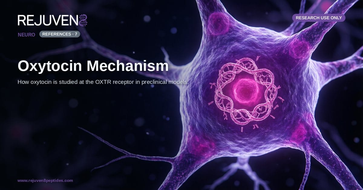
Oxytocin is a nine-amino-acid cyclic peptide that occupies a central place in the study of neuroendocrine signaling and social behavior. In preclinical literature the oxytocin mechanism is examined at the level of a single receptor, the oxytocin receptor (OXTR), a G-protein-coupled receptor expressed in defined neuronal populations of the rodent and mammalian brain. Investigations of this system ask how a small hypothalamic peptide binds its receptor, recruits an intracellular signaling cascade, and becomes associated with measurable readouts in laboratory models of social recognition, affiliation, and parental care.
The purpose of this overview is to describe how researchers characterize OXTR signaling in cell-based and animal studies. The material is presented for research use only, not for human consumption, and it makes no claim about clinical or behavioral outcomes in people. The scope is deliberately mechanistic: the structure of the peptide, the receptor it engages, the second-messenger pathways it activates, and the preclinical social-behavior and neuroendocrine paradigms in which those pathways are investigated. Oxytocin studied as a laboratory reference compound is described on the Oxytocin research listing.
The Oxytocin Molecule and the OXTR Receptor
Understanding the oxytocin mechanism begins with the physical properties of the peptide and the architecture of the receptor that recognizes it. Both are well characterized in structural and molecular studies, which makes the system a useful reference model for GPCR pharmacology.
Peptide identity
Oxytocin is a nonapeptide, a chain of nine amino acids arranged as Cys-Tyr-Ile-Gln-Asn-Cys-Pro-Leu-Gly-NH2. An intramolecular disulfide bridge between the two cysteine residues (positions 1 and 6) closes a six-residue ring and leaves a short three-residue tail. This compact, constrained shape is central to how the molecule fits its binding pocket, and it is nearly identical to the structure of vasopressin, from which oxytocin differs at only two positions. The peptide is synthesized primarily in magnocellular and parvocellular neurons of the hypothalamic paraventricular and supraoptic nuclei, then transported along axonal projections for release.
The values below reflect commonly reported laboratory attributes and are provided for comparison only.
| Attribute | Reported value |
|---|---|
| Peptide class | Cyclic nonapeptide (9 residues) |
| Amino-acid sequence | Cys-Tyr-Ile-Gln-Asn-Cys-Pro-Leu-Gly-NH2 |
| Approximate molecular weight | ~1007 Da |
| Key structural feature | Disulfide bridge (Cys1 to Cys6) |
| Primary synthesis site | Hypothalamus (paraventricular and supraoptic nuclei) |
| Cognate receptor | OXTR (oxytocin receptor) |
| Receptor class | Class A (rhodopsin-like) GPCR |
| Primary G-protein coupling | Gq/11 |
The receptor
OXTR is a class A (rhodopsin-like) G-protein-coupled receptor, a seven-transmembrane protein that translates an extracellular binding event into an intracellular response. In rodent brain tissue OXTR is mapped to discrete regions, including parts of the amygdala, nucleus accumbens, hippocampus, and cortex, and the distribution of receptor density is itself a major variable in comparative social-behavior research. Receptor availability, rather than peptide concentration alone, is frequently treated as a rate-limiting factor that shapes how a given circuit responds.
OXTR Signal Transduction
Once oxytocin occupies OXTR, the receptor initiates a defined chain of intracellular events. The canonical pathway is one of the most reproduced observations in the field and provides the biochemical backbone for interpreting behavioral data.
The Gq and phospholipase C cascade
OXTR couples primarily to Gq/11 proteins. Activation stimulates phospholipase C beta, which cleaves the membrane lipid PIP2 into two second messengers: inositol trisphosphate (IP3) and diacylglycerol (DAG). IP3 triggers release of calcium from intracellular stores, while DAG activates protein kinase C. The resulting rise in intracellular calcium is the signaling event most often quantified in cell-based assays of OXTR function, and it links receptor occupancy to downstream enzymatic and electrical changes in the neuron.
Downstream kinases and gene expression
The calcium and protein kinase C signals feed into additional cascades, including the MAPK/ERK pathway and calcium/calmodulin-dependent enzymes. In preclinical work these branches are associated with changes in transcription-factor activity, such as CREB phosphorylation, and with longer-lasting adjustments in neuronal excitability. Some studies also report context-dependent coupling of OXTR to Gi/o proteins, which illustrates that the receptor is not restricted to a single output and that cellular context influences which pathway dominates.
OXTR in Preclinical Social-Behavior Models
The reason OXTR signaling attracts sustained attention is its recurring association with social and affiliative behaviors in animal models. Several paradigms are used repeatedly across laboratories, and each provides a controlled readout of receptor function.
Social recognition in rodents
Rodents rely on olfactory cues to distinguish familiar from novel individuals. Studies using receptor knockouts and region-specific manipulation have reported that OXTR signaling in the medial amygdala contributes to social recognition memory, a finding often used as a baseline assay when characterizing the pathway. These are behavioral readouts in animals and are not statements about human cognition.
Pair bonding in prairie voles
The socially monogamous prairie vole is a widely cited model for affiliative behavior. Comparative work has linked differences in OXTR distribution, particularly in reward-associated regions such as the nucleus accumbens, to partner-preference formation in these animals. The vole model illustrates how receptor localization, not peptide level alone, can shape behavioral outcomes in a species-specific way.
Maternal and neuroendocrine physiology
Beyond central behavior, oxytocin is studied for peripheral neuroendocrine roles, including smooth-muscle contraction driven by the same Gq calcium pathway. These physiological assays provide a well-defined, quantifiable measure of receptor activation that complements behavioral work and helps validate the pharmacological tools used in mechanistic research.
Oxytocin and Vasopressin: A Signaling Comparison
Oxytocin is rarely studied in isolation from its structural sibling, vasopressin. The two nonapeptides differ by only two amino acids yet engage overlapping receptor families, and cross-reactivity between them is a persistent methodological consideration in OXTR research.
The values below reflect commonly reported laboratory attributes and are provided for comparison only.
| Feature | Oxytocin | Vasopressin (AVP) |
|---|---|---|
| Residue count | 9 | 9 |
| Approximate molecular weight | ~1007 Da | ~1084 Da |
| Residues at positions 3 and 8 | Ile, Leu | Phe, Arg |
| Primary receptor studied | OXTR | V1a, V1b, V2 |
| Primary coupling (OXTR / V1a) | Gq/11 | Gq/11 |
| Common preclinical focus | Social recognition, affiliation | Territorial behavior, fluid balance |
Because oxytocin can bind vasopressin receptors and vice versa, selective agonists, antagonists, and receptor knockouts are standard tools for isolating true OXTR-mediated effects. This is one reason well-characterized, high-purity reference material matters in mechanistic studies. Documentation such as a certificate of analysis and independent testing certificates lets researchers confirm identity and purity before an experiment. Related neuropeptides studied in adjacent behavioral and cognitive paradigms, such as Selank and Semax, are frequently examined alongside oxytocin in the broader neuropeptide literature.
Frequently Asked Questions
What is the oxytocin mechanism of action at the receptor level?
In molecular terms, oxytocin binds OXTR, a class A GPCR that couples mainly to Gq/11 proteins. This activates phospholipase C, generates IP3 and DAG, and raises intracellular calcium, which drives downstream kinase signaling. All of this is described in cell-based and animal research and does not represent a human therapeutic mechanism.
What type of receptor is OXTR?
OXTR is a seven-transmembrane, rhodopsin-like (class A) G-protein-coupled receptor. It is expressed in defined brain regions in rodent models as well as in peripheral tissues, and its regional density is a key variable in comparative studies of social behavior.
How is oxytocin signaling studied in animal models?
Researchers use receptor knockouts, selective agonists and antagonists, calcium-imaging assays, and behavioral paradigms such as social recognition and prairie-vole partner preference. These approaches isolate OXTR-specific effects and are conducted entirely in preclinical, non-human systems.
How does oxytocin differ from vasopressin?
Both are nine-residue cyclic peptides that differ at only two positions. Oxytocin is studied mainly at OXTR, while vasopressin acts at V1a, V1b, and V2 receptors. Their structural similarity produces measurable cross-reactivity, which researchers control for with selective tools.
Why is oxytocin labeled research use only?
This oxytocin is supplied strictly as a laboratory reference compound. It is provided for research use only, not for human consumption, and no dosing, safety, or efficacy claims are made. Its scientific interest lies in the mechanistic questions described above.
What documentation matters when sourcing oxytocin for research?
Identity and purity data are essential for reproducible mechanistic work. A certificate of analysis, mass-spectrometry identity confirmation, and independent testing help ensure that observed effects reflect the intended peptide. Guidance on choosing a research peptide supplier outlines what to check.
References and Further Reading
- Gimpl G and Fahrenholz F (2001). The oxytocin receptor system: structure, function, and regulation. Physiological Reviews. PubMed: oxytocin receptor system structure function
- Jurek B and Neumann ID (2018). The oxytocin receptor: from intracellular signaling to behavior. Physiological Reviews. PubMed: oxytocin receptor intracellular signaling behavior
- Young LJ and Wang Z. The neurobiology of pair bonding in rodent models. PubMed: oxytocin pair bonding prairie vole
- Insel TR. Oxytocin and the neurobiology of social behavior. PubMed: oxytocin social behavior neurobiology
- Carter CS. Oxytocin, vasopressin, and social bonding in animal models. PubMed: oxytocin vasopressin social bonding
- Stoop R. Neural circuits and signaling of the oxytocin system. PubMed: oxytocin neural circuits signaling
- Grinevich V and Neumann ID. Oxytocin neuron activity and central release. PubMed: oxytocin neuron central release