Research Use Only. The information presented here is for scientific and educational purposes. These compounds are not intended for human consumption, self-administration, or therapeutic use.
Introduction
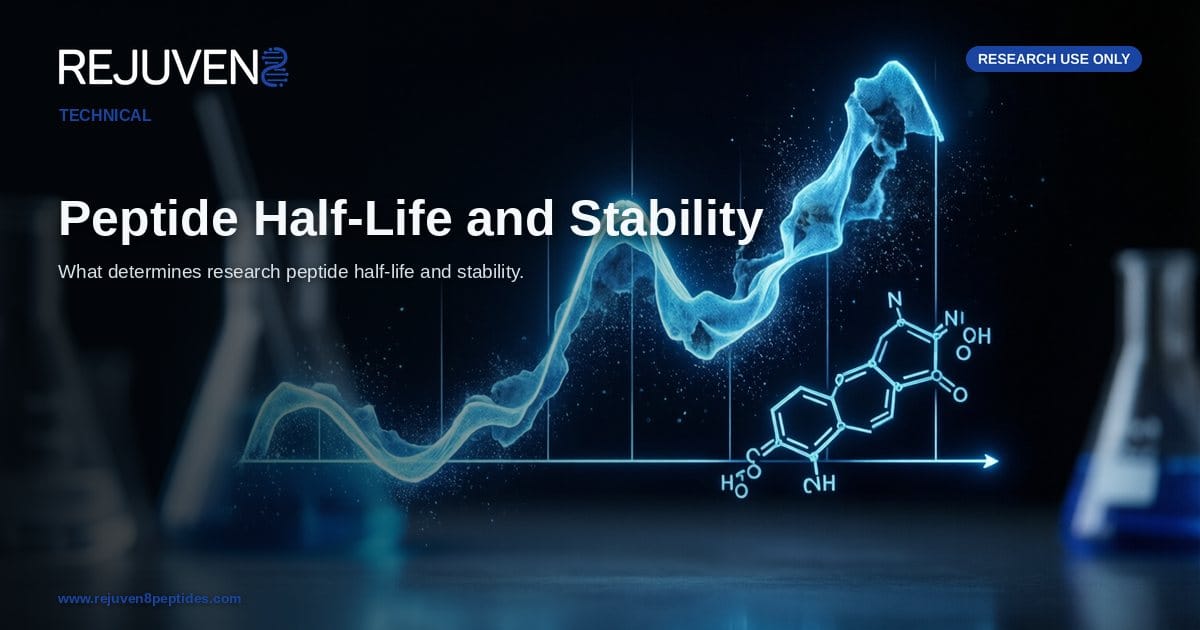
Peptide half-life and stability are among the most studied physicochemical and pharmacokinetic parameters in preclinical peptide science. Half-life describes the time required for the concentration of a peptide in circulation to fall by half within a model system, and it is governed largely by three processes: enzymatic cleavage by proteolytic enzymes, removal through renal clearance, and the extent to which a molecule binds carrier proteins such as serum albumin. These pathways define how long a research peptide remains measurable during an in-vitro assay or an animal study.
Stability and half-life are related but distinct ideas. Chemical stability refers to how well a peptide resists breakdown in a vial or buffer, while biological half-life reflects how quickly a living system degrades and eliminates it. This article surveys the determinants scientists measure and manipulate: sequence features, structural modifications, and storage conditions. All material discussed here is handled for research use only, not for human consumption, and every attribute is framed in terms of laboratory investigation rather than any clinical outcome.
What Peptide Half-Life Describes
Plasma Half-Life as a Pharmacokinetic Measure
Plasma half-life (often written as t-half) is the interval over which the measured concentration of a peptide in the circulation of a model organism decreases by fifty percent. It is derived from concentration-versus-time curves generated in animal studies or perfusion models. A short half-life (minutes) indicates rapid degradation or elimination, while a long half-life (hours or days) points to strong resistance to clearance. Because most native sequences are cleared quickly, research attention focuses on the structural and formulation factors that lengthen persistence.
In-Vitro Stability Compared With In-Vivo Half-Life
A recurring theme in stability research is the gap between in-vitro stability and in-vivo half-life. In-vitro work examines how a peptide holds up in a controlled environment such as buffer, serum incubations, or storage. In-vivo half-life captures the additional forces present in a living system: active enzymes in plasma and tissue, distribution into compartments, and clearance organs such as the kidney and liver. A peptide can be stable in a sealed vial yet still show a short in-vivo half-life once enzymes and filtration act on it. For this reason researchers report both measures, and a certificate of analysis that documents purity is a useful starting point, as explained in the COA and purity guide.
Why Peptides Degrade: Proteases and Clearance
Native peptides show short half-lives because the body is highly efficient at dismantling them, mainly through enzymatic proteolysis and renal filtration.
Proteolytic Enzymes
Proteases (also called peptidases) are enzymes that hydrolyze the peptide bonds linking amino acids. Exopeptidases trim residues from the ends of a chain, and dipeptidyl peptidase-4 (DPP-4) is a well-studied example that cleaves the N-terminus of many regulatory peptides. Endopeptidases cut internal bonds within the sequence. Because these enzymes recognize specific residues and bond geometries, amino acid composition and backbone accessibility strongly influence how fast degradation proceeds. Peptides with exposed, protease-favored cleavage sites break down within minutes in serum studies, whereas sequences that shield those sites persist longer.
Renal Filtration and Molecular Size
The second major route is renal clearance. The glomerulus filters small molecules from the blood, and peptides below roughly the low kilodalton range are removed readily. As a result, molecular weight and effective hydrodynamic size are meaningful determinants of half-life: larger or protein-bound constructs are filtered more slowly. Several extension strategies therefore aim to make a peptide behave as though it were larger. The table below compares commonly studied research peptides by class, size, and structure.
The values below reflect commonly reported laboratory attributes and are provided for comparison only.
| Research peptide | Class | Approx. molecular weight | Reported half-life character |
|---|---|---|---|
| Ipamorelin | GH secretagogue pentapeptide (5 aa) | ~712 Da | Short, reported on the order of minutes |
| Sermorelin | GHRH analog (residues 1-29) | ~3358 Da | Short, rapid peptidase cleavage in studies |
| CJC-1295 with DAC | GHRH analog plus drug affinity complex | ~3649 Da | Markedly extended via albumin binding |
| BPC-157 | Synthetic pentadecapeptide (15 aa) | ~1419 Da | Noted chemical robustness in buffer studies |
| GLP-1 SM | Acylated GLP-1 receptor analog | ~4114 Da | Extended via fatty-acid acylation |
The contrast between short-lived analogs such as Sermorelin and the extended profile of CJC-1295 with DAC shows how a single structural addition reshapes pharmacokinetic behavior in research models.
Structural Modifications That Extend Half-Life
Because native peptides are cleared so quickly, medicinal-chemistry research has produced a toolkit of deliberate modifications, each targeting one of the degradation routes above: resisting proteases, slowing filtration, or promoting protein binding.
Albumin Binding Through DAC and Acylation
Two of the most influential approaches exploit serum albumin, the abundant carrier protein in blood. The drug affinity complex (DAC) attaches a group that forms a reversible covalent bond with albumin, parking the peptide on a large, long-circulating protein. Fatty-acid acylation achieves a similar result non-covalently, adding a lipid chain that inserts into an albumin binding pocket. In both cases the bound peptide is shielded from enzymatic cleavage and is too large for rapid renal filtration, so reported half-life values in animal studies rise from minutes toward hours or days. CJC-1295 with DAC is a widely cited example of the covalent approach, while acylated analogs in the GLP-1 SM class illustrate the fatty-acid route.
Backbone, Terminal, and Size Modifications
Other strategies alter the peptide itself rather than recruiting a carrier. D-amino acid substitution replaces natural L-residues with their mirror-image form, which many stereospecific proteases fail to recognize. Cyclization constrains the backbone into a ring that hinders exopeptidase access. N-terminal and C-terminal capping, such as acetylation or amidation, blocks the entry points that trimming enzymes use. PEGylation attaches polyethylene glycol chains that increase hydrodynamic size and slow renal filtration.
The entries below reflect commonly reported laboratory attributes and are provided for comparison only.
| Strategy | Mechanism | Reported effect | Research example |
|---|---|---|---|
| Drug affinity complex (DAC) | Reversible covalent binding to albumin | Substantial extension | CJC-1295 with DAC |
| Fatty-acid acylation | Non-covalent albumin binding via lipid chain | Substantial extension | Acylated GLP-1 SM analog |
| D-amino acid substitution | Resists stereospecific protease cleavage | Moderate extension | Various GHRH and melanocortin analogs |
| Cyclization | Constrains backbone against exopeptidases | Moderate extension | Cyclic research peptides |
| Terminal capping | Blocks N-terminal and C-terminal trimming | Moderate extension | Acetylated or amidated analogs |
| PEGylation | Adds hydrodynamic size, slows filtration | Substantial extension | PEGylated peptide constructs |
Storage and Handling Stability
A peptide must survive storage and preparation before any assay begins, so handling stability is where researchers exert the most direct control over the same chemistry.
Lyophilized Versus Reconstituted Material
Most research peptides are supplied as a lyophilized (freeze-dried) powder because removing water dramatically slows the hydrolysis and oxidation reactions that degrade a sequence. In dry form, kept cold and sealed, many peptides are reported to remain stable for extended periods. Once reconstituted into solution, the same molecule becomes far more vulnerable, since water, dissolved oxygen, and residual enzymes can act on it, so it is generally refrigerated and used within a defined window. Solvent choice and mixing technique are covered in the bacteriostatic water reconstitution guide, which this article does not restate at length.
Temperature, Light, and Freeze-Thaw
Several environmental factors accelerate breakdown. Elevated temperature speeds every degradation reaction, which is why cold storage is standard. Light exposure can drive photo-oxidation in sensitive residues, so amber vials and dark storage are common precautions. Repeated freeze-thaw cycles are a frequent cause of loss, because ice formation and concentration changes during thawing can fragment or aggregate a peptide; single-use aliquots minimize this stress. Solution pH and buffer composition matter as well, since some sequences are stable only within a narrow range. Sourcing well-characterized material with documented purity supports reproducible work, as outlined in the supplier selection guide alongside the published certificates of analysis.
Frequently Asked Questions
What does peptide half-life mean in research?
In research, peptide half-life refers to the time required for the plasma concentration of a compound to decrease by half after administration in a model system. It is a pharmacokinetic parameter measured in vitro or in animal studies, and it reflects the combined effects of enzymatic degradation, renal clearance, and plasma protein binding.
What determines how long a peptide lasts in the body?
In preclinical models, persistence is governed mainly by proteolytic enzymes that cleave peptide bonds, by renal filtration that removes small molecules, and by the degree to which a peptide binds carrier proteins such as albumin. Structural features including amino acid composition, sequence length, and terminal chemistry all influence how quickly these processes act.
What is the difference between in-vitro stability and in-vivo half-life?
In-vitro stability describes how well a peptide resists chemical breakdown in a controlled setting such as buffer, serum, or storage conditions. In-vivo half-life describes how long the compound remains measurable in a living model system, where circulation, enzymes, and clearance organs all contribute. A peptide can be chemically stable in a vial yet still show a short in-vivo half-life.
How do modifications like DAC and acylation extend peptide half-life?
The drug affinity complex (DAC) and fatty-acid acylation both work by promoting binding to serum albumin. An albumin-bound peptide is shielded from rapid enzymatic cleavage and renal filtration, so reported half-life values in animal studies increase substantially. CJC-1295 with DAC is a commonly cited research example of this strategy.
How should research peptides be stored to preserve stability?
Lyophilized (freeze-dried) peptides are generally reported as most stable when kept cold, dry, and protected from light. After reconstitution, material is typically refrigerated and protected from repeated freeze-thaw cycles, which can degrade sensitive sequences. These handling practices apply to research use only, not for human consumption.
Does molecular weight affect peptide half-life?
Molecular size influences renal clearance, since the kidneys filter small peptides more readily than larger constructs. Strategies that increase effective hydrodynamic size, such as PEGylation or albumin binding, tend to slow filtration and lengthen reported half-life. Molecular weight alone does not determine stability, because enzymatic susceptibility also depends on sequence and structure.