Research Use Only. The information presented here is for scientific and educational purposes. These compounds are not intended for human consumption, self-administration, or therapeutic use.
Introduction
Insulin-like growth factor 1 (IGF-1) is one of the most studied anabolic signaling peptides in cell biology, sitting downstream of growth hormone on the somatotropic axis and acting through the IGF-1 receptor (IGF-1R) to influence proliferation, differentiation, and survival in cultured cells. Native IGF-1 is a 70-amino-acid polypeptide that circulates almost entirely bound to a family of IGF binding proteins (IGFBPs), a feature that shapes both how long it persists and how much of it is free to engage its receptor. For laboratories modeling the growth-hormone axis in vitro, that binding behavior is central to interpreting any result.
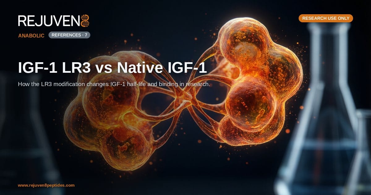
IGF-1 LR3 (Long R3 IGF-1) is an engineered analog created specifically to change that behavior. By combining an amino-acid substitution at position 3 with a 13-residue N-terminal extension, the LR3 variant sharply lowers its affinity for IGFBPs while retaining activity at IGF-1R. This article compares native IGF-1 and IGF-1 LR3 across the three axes that matter most in laboratory work: the Long R3 structural modification, IGFBP binding, and reported half-life. All discussion here is framed for research use only, not for human consumption.
Native IGF-1: The Baseline Molecule
Native IGF-1, historically called somatomedin C, is a single-chain polypeptide of 70 amino acids with an approximate molecular weight of 7,649 Da. Its fold is stabilized by three intramolecular disulfide bonds and is structurally homologous to proinsulin, which is why IGF-1 shows weak cross-reactivity at the insulin receptor. In classic endocrine models, hepatic IGF-1 is produced in response to pituitary growth hormone and mediates many of the downstream growth-promoting signals attributed to GH.
Receptor and Signaling
IGF-1 binds the IGF-1 receptor, a transmembrane receptor tyrosine kinase. Receptor engagement triggers autophosphorylation and recruitment of adaptor proteins, activating the PI3K/Akt and Ras/MAPK cascades that serve as common readouts in proliferation and cell-survival assays. Because native IGF-1 and its analogs share this receptor, comparisons between them usually hold receptor signaling roughly constant and focus instead on availability, meaning how much peptide actually reaches the receptor.
The IGFBP Problem
In circulation and in many culture systems, roughly 99 percent of native IGF-1 is bound to one of six IGF binding proteins, predominantly IGFBP-3, often in a ternary complex with the acid-labile subunit (ALS). This binding protects IGF-1 from clearance but also sequesters it, keeping free peptide low. Free native IGF-1 has a very short reported half-life on the order of ten minutes, while the protected ternary complex extends persistence to many hours. That split makes native IGF-1 behavior difficult to standardize when IGFBP levels vary between experimental setups.
The Long R3 Modification Explained
IGF-1 LR3 addresses the IGFBP problem through two deliberate structural edits. Together they give the molecule its full name, Long R3 IGF-1, and its distinct behavior in research models.
The R3 Substitution
The “R3” in the name refers to the replacement of glutamic acid with arginine at position 3 of the mature IGF-1 sequence. This single substitution sharply reduces the analog’s affinity for IGFBPs. With less peptide captured by binding proteins, a larger fraction remains free to interact with IGF-1R, which is the practical reason the analog appears more potent in cell culture than an equivalent mass of native IGF-1.
The “Long” N-Terminal Extension
The “Long” prefix denotes a 13-amino-acid extension peptide added to the N-terminus of the molecule. This extension increases the overall size of the analog to 83 amino acids and an approximate molecular weight of 9,111 Da. The combined effect of the extension and the Arg3 substitution is an analog that resists IGFBP sequestration and, in reported research, shows a substantially longer functional half-life than free native IGF-1. The lab-tested IGF-1 LR3 research peptide is supplied as this modified 83-residue form.
IGF-1 LR3 vs Native IGF-1: Side by Side
The table below summarizes the structural and behavioral differences most often cited when researchers choose between the two molecules. These values reflect commonly reported laboratory attributes and are provided for comparison only.
| Attribute | Native IGF-1 | IGF-1 LR3 |
|---|---|---|
| Class | Endogenous growth factor peptide | Engineered IGF-1 analog |
| Sequence length | 70 amino acids | 83 amino acids |
| Approx. molecular weight | ~7,649 Da | ~9,111 Da |
| Key modification | None (wild-type sequence) | Arg3 substitution plus 13-residue N-terminal extension |
| IGFBP affinity | High (mostly bound) | Markedly reduced |
| Free fraction in culture | Low | High |
| Reported half-life | Very short when free (~minutes) | Substantially longer |
| Primary receptor | IGF-1R | IGF-1R |
The headline takeaway is that both molecules act at the same receptor, but IGF-1 LR3 changes the delivery problem. By evading IGFBPs, it keeps more peptide free for longer, which is why it is frequently selected as a research tool for studying sustained IGF-1R signaling. Laboratories comparing anabolic peptides often place it alongside other growth-axis compounds, and the broader recovery and tissue-repair peptide overview provides that wider context.
Half-Life and Binding in Research Models
The most consequential practical difference between the two molecules is how binding-protein affinity translates into duration of action. Because native IGF-1 is heavily bound, its free concentration is buffered by IGFBP levels that can shift with the culture medium, serum content, or tissue source. IGF-1 LR3, with its reduced IGFBP binding, is less sensitive to that buffering, giving more reproducible free-peptide exposure across conditions.
Why Half-Life Matters in Vitro
A longer functional half-life means the analog can sustain receptor stimulation over an extended window without repeated re-dosing of the culture, a convenience that has made LR3 a common choice in proliferation and differentiation studies. Researchers should note that reported half-life figures vary with the model system and are not directly transferable between in-vitro and in-vivo contexts. The comparison is therefore best read as a difference in binding behavior rather than a fixed numeric ratio.
Handling and Documentation
As with any research peptide, reconstitution, cold storage, and minimizing freeze-thaw cycles influence the integrity of both molecules; the bacteriostatic water reconstitution guide covers the general handling steps. Purchasers should also verify identity and purity against a certificate of analysis, and the COA purity primer explains how to read one. These checks matter especially for an engineered analog, where sequence-length and molecular-weight confirmation are what distinguish it from the wild-type peptide. Both molecules can be found among the anabolic options in the wider research peptide catalog.
Frequently Asked Questions
What is the main difference between IGF-1 LR3 and native IGF-1?
Native IGF-1 is the wild-type 70-amino-acid growth factor that binds tightly to IGF binding proteins, while IGF-1 LR3 is an 83-amino-acid analog engineered with an arginine substitution at position 3 and a 13-residue N-terminal extension that together reduce IGFBP binding and extend its reported functional half-life in research models.
What does Long R3 actually mean?
The R3 portion refers to arginine (R) replacing glutamic acid at position 3 of the IGF-1 sequence, and Long refers to the 13-amino-acid extension added to the N-terminus. Combined, these two edits define the Long R3 IGF-1 analog.
Why does IGF-1 LR3 have a longer half-life?
The Arg3 substitution lowers the analog’s affinity for IGF binding proteins. Because much of native IGF-1 rapid sequestration and turnover is tied to IGFBP interactions, reducing that binding leaves more free analog available for longer in reported research models.
Do IGF-1 LR3 and native IGF-1 act on the same receptor?
Yes. Both engage the IGF-1 receptor (IGF-1R), a receptor tyrosine kinase that activates the PI3K/Akt and MAPK signaling pathways. The modifications in LR3 change availability and binding-protein behavior rather than the receptor target itself.
Is IGF-1 LR3 more potent than native IGF-1?
In cell-culture systems, IGF-1 LR3 often appears more potent per unit mass because it is not sequestered by IGFBPs, leaving a larger free fraction to stimulate the receptor. Potency comparisons depend heavily on the assay and on the IGFBP content of the system being used.
How are these compounds intended to be used?
Both are supplied strictly for research use only, not for human consumption. They are laboratory reference materials for in-vitro and preclinical study of the IGF-1 axis, not products for self-administration or therapeutic use.
References and Further Reading
- Rinderknecht E, Humbel RE (1978), determination of the primary amino-acid sequence of human IGF-1. PubMed: human insulin-like growth factor I amino acid sequence
- Francis, Ballard and colleagues, characterization of Long R3 IGF-1 analogs and their reduced binding-protein affinity. PubMed: Long R3 IGF-I analogue
- Firth SM, Baxter RC, cellular actions of the IGF binding proteins. PubMed: insulin-like growth factor binding proteins
- Jones JI, Clemmons DR, review of insulin-like growth factors and their binding proteins. PubMed: insulin-like growth factors and their binding proteins
- IGF-1 receptor signaling through the PI3K/Akt and MAPK pathways. PubMed: IGF-1 receptor signaling
- IGF-1 ternary complex with IGFBP-3 and the acid-labile subunit and its effect on persistence. PubMed: IGF-1 ternary complex acid-labile subunit
- Comparative potency of Long R3 IGF-1 in cell proliferation assays. PubMed: Long R3 IGF-I cell proliferation