Research Use Only. The information presented here is for scientific and educational purposes. These compounds are not intended for human consumption, self-administration, or therapeutic use.
Introduction
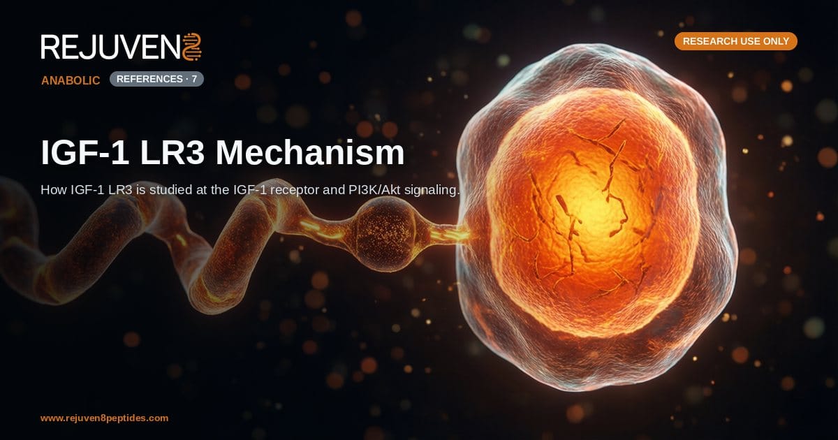
IGF-1 LR3, or Long R3 Insulin-like Growth Factor 1, is a recombinant analog of the endogenous peptide hormone insulin-like growth factor 1 (IGF-1). In laboratory research it is studied as a tool for probing signaling through the IGF-1 receptor (IGF-1R), a receptor tyrosine kinase that sits at the center of anabolic and growth-related pathways. The word “mechanism” in the context of IGF-1 LR3 refers to how the modified peptide engages this receptor and the intracellular cascades, principally the PI3K/Akt axis, that follow receptor activation in cell and animal models.
This article reviews what is known about the structure of IGF-1 LR3, how its two defining modifications alter its behavior relative to native IGF-1, and the receptor-level events that make it a subject of preclinical interest. All of the material below is framed for research use only, not for human consumption. Nothing here describes clinical use, dosing, or therapeutic outcomes; the goal is to summarize the studied molecular biology of the compound for scientific and educational purposes.
What Is IGF-1 LR3?
IGF-1 LR3 is a single-chain polypeptide engineered from the sequence of human IGF-1. Native IGF-1 is a 70-amino-acid peptide of roughly 7,649 Da that circulates as a mediator of growth hormone (GH) signaling. IGF-1 LR3 carries two deliberate changes: a substitution of arginine for glutamic acid at position 3 (the “R3” designation), and a 13-amino-acid extension added to the N-terminus (the “Long” designation). Together these modifications extend the molecule to 83 amino acids and an approximate molecular weight near 9,100 Da.
Structural modifications
The Arg3 substitution and the N-terminal extension are not arbitrary. In research characterizations, both changes reduce the affinity of the peptide for the insulin-like growth factor binding proteins (IGFBPs), the carrier proteins that normally sequester circulating IGF-1. This is the single feature most often cited when investigators explain why the analog behaves differently from the parent molecule in vitro.
The values in the table below reflect commonly reported laboratory attributes and are provided for comparison only.
| Property | Reported value |
|---|---|
| Common name | IGF-1 LR3 (Long R3 IGF-1) |
| Class | Recombinant IGF-1 analog, single-chain polypeptide |
| Sequence length | 83 amino acids |
| Approx. molecular weight | ~9,100 Da |
| Key modifications | Arg3 substitution plus 13-residue N-terminal extension |
| Primary studied target | IGF-1 receptor (IGF-1R) |
| Downstream pathways studied | PI3K/Akt/mTOR and MAPK/ERK |
As a recombinant research peptide, IGF-1 LR3 is typically supplied as a lyophilized powder for laboratory handling; general reconstitution practice is covered in the bacteriostatic water reconstitution guide rather than restated here.
The IGF-1 Receptor and Downstream Signaling
The primary studied target of IGF-1 LR3 is the IGF-1 receptor (IGF-1R), a transmembrane receptor tyrosine kinase structurally related to the insulin receptor. IGF-1R exists as a pre-formed dimer; ligand binding to the extracellular alpha subunits drives a conformational change that activates the intracellular beta-subunit kinase domains.
Receptor autophosphorylation
When IGF-1 or an analog such as IGF-1 LR3 engages IGF-1R, the receptor undergoes autophosphorylation on tyrosine residues. This creates docking sites for adaptor proteins, most notably the insulin receptor substrate (IRS) family and Shc. These adaptors are the branch point from which the two best-characterized IGF-1R pathways diverge.
PI3K/Akt and the anabolic branch
Recruitment of IRS proteins activates phosphoinositide 3-kinase (PI3K), which generates the lipid second messenger PIP3 and in turn activates the serine/threonine kinase Akt (also called protein kinase B). Akt is a central node in what researchers describe as anabolic signaling: it influences the mechanistic target of rapamycin (mTOR), which is associated in preclinical models with protein synthesis, and it modulates factors linked to cell survival and glucose uptake. The PI3K/Akt/mTOR axis is the pathway most frequently invoked when the anabolic character of IGF-1 signaling is discussed in the literature.
The MAPK/ERK branch
A parallel branch runs through Shc, Grb2, and Ras to the mitogen-activated protein kinase (MAPK) cascade, culminating in ERK1/2 activation. This route is more often associated in research with proliferation and gene transcription than with the metabolic events attributed to the Akt branch. IGF-1 LR3, by virtue of engaging the same receptor, is studied as a ligand that can drive both branches.
Reduced IGFBP Binding and the LR3 Modification
In serum-containing culture and in circulation, most IGF-1 is bound to one of six IGF binding proteins (IGFBPs), which regulate its availability and half-life. The defining research rationale for IGF-1 LR3 is that its structural modifications markedly reduce this binding. With less of the peptide captured by IGFBPs, a greater fraction remains free to interact with IGF-1R in experimental systems, and the effective duration of receptor engagement is reported to be longer than that of native IGF-1.
The values below reflect commonly reported laboratory attributes and are provided for comparison only.
| Attribute | Native IGF-1 | IGF-1 LR3 |
|---|---|---|
| Sequence length | 70 amino acids | 83 amino acids |
| Approx. molecular weight | ~7,649 Da | ~9,100 Da |
| Residue at position 3 | Glutamic acid (Glu) | Arginine (Arg) |
| N-terminal extension | None | 13-residue extension |
| IGFBP binding affinity | High | Markedly reduced |
| Relative research half-life | Shorter | Longer |
| Class | Endogenous peptide hormone | Recombinant analog |
Why the comparison matters
For researchers, the reduced IGFBP affinity is what makes IGF-1 LR3 a convenient probe: it allows receptor-level questions to be studied with less confounding from binding-protein dynamics. This is a molecular-biology rationale for the analog’s use in the laboratory, not a statement of any human or veterinary application.
Anabolic Signaling in Preclinical Research
Because IGF-1 LR3 activates IGF-1R robustly in cell culture, it appears frequently in in-vitro studies of myoblast and other cell-line behavior, and in animal models examining the GH/IGF-1 axis. IGF-1 sits downstream of growth hormone in that axis, which is why the compound is often discussed alongside GH secretagogues such as CJC-1295 and Ipamorelin in the broader research context.
Position in the GH/IGF-1 axis
Growth hormone released from the pituitary stimulates hepatic and local production of IGF-1, which then acts on IGF-1R. Analogs like IGF-1 LR3 let investigators study the receptor-level arm of this axis directly, without relying on upstream GH stimulation. For a broader survey of peptides examined for tissue and recovery research, see the recovery and tissue repair overview.
Handling and verification
As with any research peptide, purity and identity matter for reproducible signaling studies. Reviewing a certificate of analysis (COA) is standard practice before a compound is used at the bench. IGF-1 LR3 is offered as a lab-tested research peptide for these purposes. This material is intended for research use only, not for human consumption.
Frequently Asked Questions
What is the mechanism of action of IGF-1 LR3?
In research settings, IGF-1 LR3 is studied as an agonist of the IGF-1 receptor (IGF-1R). Binding activates the receptor’s tyrosine kinase domain, triggering autophosphorylation and the recruitment of IRS adaptor proteins. This leads to activation of the PI3K/Akt/mTOR pathway associated with anabolic signaling and, in parallel, the MAPK/ERK pathway associated with proliferation.
How does IGF-1 LR3 differ from native IGF-1?
IGF-1 LR3 carries an arginine-for-glutamic-acid substitution at position 3 and a 13-amino-acid N-terminal extension. These changes lengthen the peptide to 83 amino acids (about 9,100 Da, versus roughly 7,649 Da for native IGF-1) and, most importantly for research, markedly reduce its binding to the IGF binding proteins.
What is the role of the IGF-1 receptor in IGF-1 LR3 signaling?
IGF-1R is the transmembrane receptor tyrosine kinase that IGF-1 LR3 is studied to activate. It is the entry point for all downstream signaling: without receptor engagement and autophosphorylation, neither the PI3K/Akt branch nor the MAPK/ERK branch is initiated.
Why does IGF-1 LR3 have reduced IGFBP binding?
The two structural modifications, particularly the charge change introduced by the Arg3 substitution and the steric contribution of the N-terminal extension, lower the peptide’s affinity for the IGFBP carrier proteins. In experimental systems this leaves more of the peptide free to reach IGF-1R.
What is the PI3K/Akt pathway in IGF-1 research?
PI3K/Akt is the intracellular signaling cascade most associated with the anabolic events attributed to IGF-1R activation. PI3K produces the lipid messenger PIP3, which activates Akt; Akt then influences mTOR and other targets linked in preclinical models to protein synthesis and cell survival.
Is IGF-1 LR3 intended for human use?
No. IGF-1 LR3 is a research compound offered for research use only, not for human consumption. The information here describes its studied molecular biology in preclinical and in-vitro contexts and does not constitute any therapeutic, dosing, or clinical guidance.
References and Further Reading
- Rinderknecht E, Humbel RE. Foundational characterization of the primary structure of insulin-like growth factor 1 (1978). PubMed: insulin-like growth factor 1 primary structure
- Reviews of IGF-1 receptor signaling and its role in growth and metabolism. PubMed: IGF-1 receptor signaling
- Literature on the IGF-1 receptor as a tyrosine kinase and its autophosphorylation. PubMed: IGF-1 receptor tyrosine kinase
- PI3K/Akt signaling as a mediator of anabolic and survival responses. PubMed: PI3K Akt signaling pathway
- Studies of the insulin-like growth factor binding proteins (IGFBPs) and their regulation of IGF availability. PubMed: IGF binding proteins
- Francis and colleagues, and later work, on Long R3 IGF-1 and related recombinant analogs with reduced IGFBP affinity. PubMed: Long R3 IGF-1 analog
- mTOR signaling downstream of Akt in the regulation of protein synthesis. PubMed: mTOR protein synthesis signaling